U-Net DFE plus ASPP Segmentation Architecture
About This Architecture
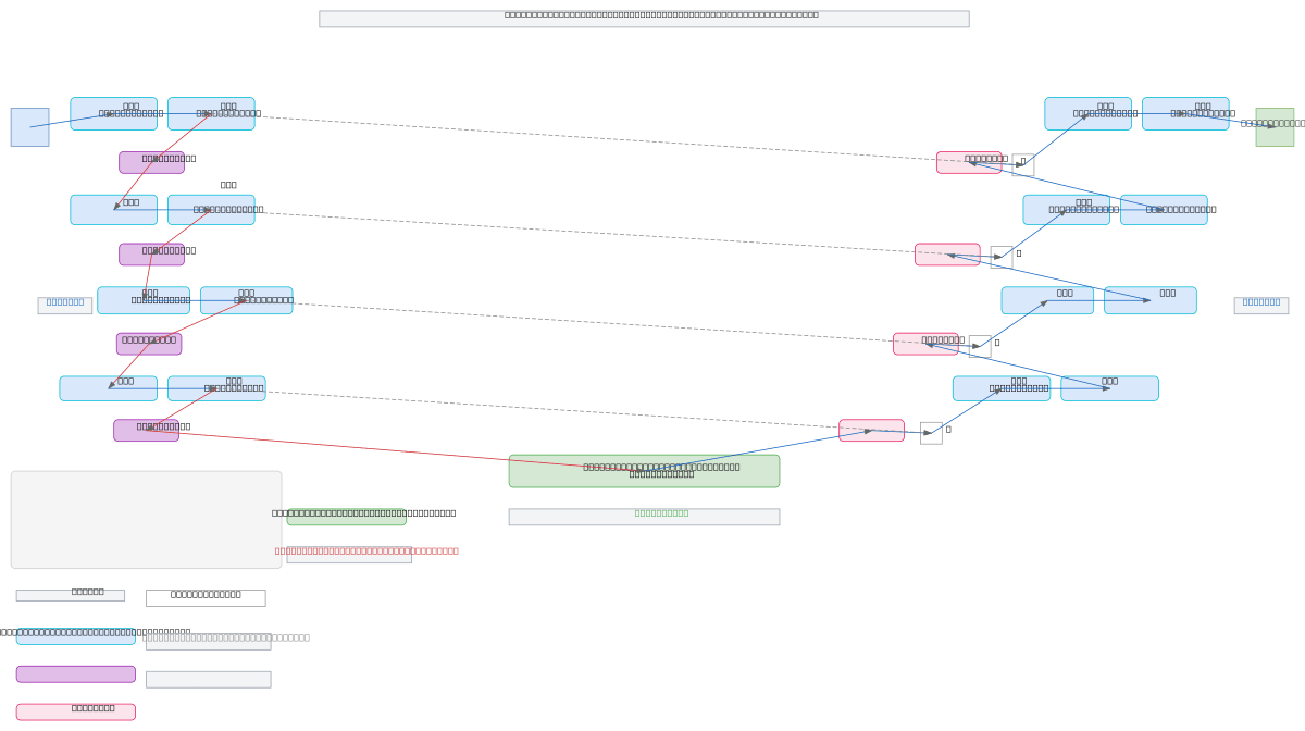
U-Net architecture enhanced with Deformable Feature Enhancement (DFE) modules and Atrous Spatial Pyramid Pooling (ASPP) for high-precision brain MRI segmentation. The encoder progressively downsamples Brain MRI Input through four DFE blocks (64ch to 512ch) with skip connections, while the ASPP bottleneck captures multi-scale contextual features at 1024ch 16x16 resolution. The decoder mirrors the encoder, upsampling through concatenated skip connections and DFE blocks to produce detailed Segmentation Output at original resolution. This architecture combines spatial adaptability via deformable convolutions with multi-scale context awareness, delivering superior performance on medical image segmentation tasks. Fork and customize this diagram on Diagrams.so to document your own U-Net variants or integrate into research papers and technical documentation.
People also ask
How does U-Net with Deformable Feature Enhancement and ASPP improve medical image segmentation?
This U-Net variant enhances standard segmentation by integrating DFE modules for spatially adaptive feature learning and ASPP for multi-scale contextual awareness at the bottleneck. The encoder progressively downsamples Brain MRI Input while skip connections preserve spatial details, enabling the decoder to reconstruct precise Segmentation Output with both local and global context.
- Domain:
- Ml Pipeline
- Audience:
- Machine learning engineers and medical imaging researchers implementing semantic segmentation models
Generated by Diagrams.so — AI architecture diagram generator with native Draw.io output. Fork this diagram, remix it, or download as .drawio, PNG, or SVG.