Oculomotor Apparatus - Muscles, Nerves, and Nuclei
About This Architecture
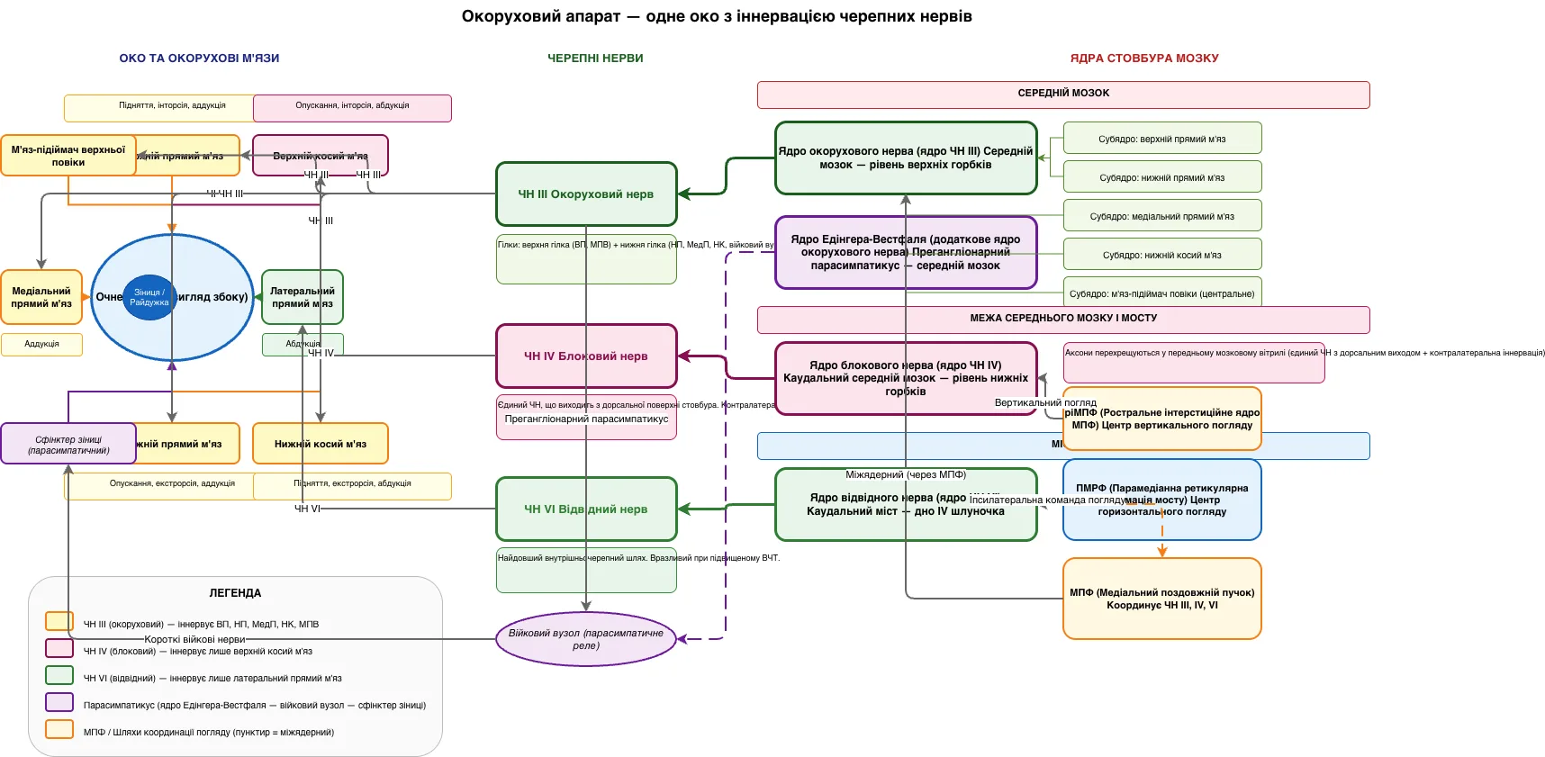
The oculomotor apparatus integrates three cranial nerves—CN III, CN IV, and CN VI—with six extraocular muscles and brainstem nuclei to coordinate eye movement and pupil function. CN III (oculomotor) innervates the superior rectus, inferior rectus, medial rectus, inferior oblique, and levator palpebrae; CN IV (trochlear) controls the superior oblique via unique dorsal brainstem exit and contralateral decussation; CN VI (abducens) drives the lateral rectus and has the longest intracranial course, making it vulnerable to raised intracranial pressure. The oculomotor nucleus resides at the midbrain level of the superior colliculus with sub-nuclei for each muscle, while the Edinger-Westphal nucleus provides parasympathetic preganglionic fibers to the ciliary ganglion for pupil and accommodation control. Horizontal gaze is coordinated by the paramedian pontine reticular formation (PPRF) in the pons, and vertical gaze by the rostral interstitial nucleus of the MLF (riMLF) in the midbrain, with the medial longitudinal fasciculus (MLF) synchronizing all three cranial nerves. This diagram is essential for understanding oculomotor palsies, internuclear ophthalmoplegia, and brainstem lesion localization in clinical neurology. Fork and customize this anatomy reference on Diagrams.so to create study guides, teaching slides, or clinical case presentations.
People also ask
What are the anatomical relationships between the oculomotor, trochlear, and abducens nerves and their brainstem nuclei?
The oculomotor nucleus (CN III) at the midbrain superior colliculus level innervates five extraocular muscles and the levator palpebrae via superior and inferior divisions. The trochlear nucleus (CN IV) at the caudal midbrain uniquely exits dorsally with contralateral decussation to innervate only the superior oblique. The abducens nucleus (CN VI) in the caudal pons innervates the lateral rectus a
- Domain:
- Other
- Audience:
- Medical students and neurology residents studying cranial nerve anatomy and oculomotor control
Generated by Diagrams.so — AI architecture diagram generator with native Draw.io output. Fork this diagram, remix it, or download as .drawio, PNG, or SVG.