MambInception - MRI Brain Tumor Classification
About This Architecture
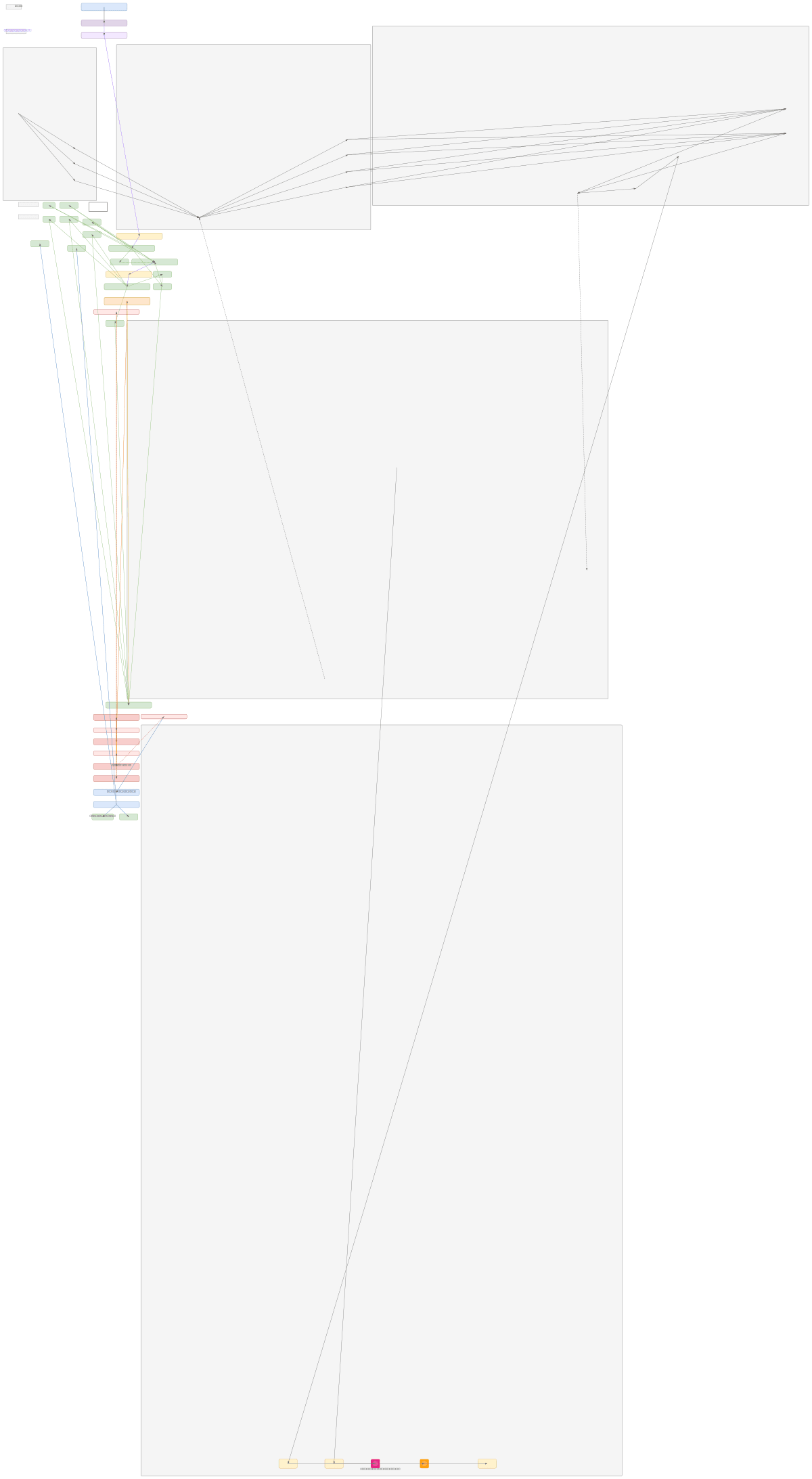
MambInception combines convolutional feature extraction with state-space sequence modeling for MRI brain tumor classification, processing 224×224 input images through Inception blocks and Mamba layers to distinguish glioma, meningioma, pituitary adenoma, and healthy tissue. The architecture flows from a stem convolution block through two Inception modules that progressively reduce spatial dimensions while expanding feature channels, then transitions to patch embedding and four Mamba blocks with selective SSM gating for efficient sequence modeling. This hybrid CNN-Mamba approach leverages OCI's compute infrastructure to achieve high accuracy on medical imaging tasks while maintaining computational efficiency through depthwise convolutions and selective attention mechanisms. Fork this diagram on Diagrams.so to customize layer configurations, adjust patch sizes, or adapt the classification head for your specific tumor dataset and OCI deployment requirements. The four-class output head with softmax normalization enables direct integration with OCI Data Science pipelines for model training and inference.
People also ask
How does MambInception combine convolutional and sequence modeling for MRI brain tumor classification?
MambInception uses Inception blocks for hierarchical feature extraction from 224×224 MRI images, progressively reducing spatial dimensions to 28×28×512, then applies patch embedding and four Mamba blocks with selective SSM gating for efficient sequence modeling. The architecture classifies tumors into four categories (glioma, meningioma, pituitary adenoma, no tumor) through a classification head e
- Domain:
- Ml Pipeline
- Audience:
- Machine learning engineers building medical imaging classification models on OCI
Generated by Diagrams.so — AI architecture diagram generator with native Draw.io output. Fork this diagram, remix it, or download as .drawio, PNG, or SVG.